Review Visualize the System Concepts & Images
Plant video: Shows the similarity of the structure of a plant’s root system to the cardiovascular system of humans.
 The first point of this comparison is seeing the big picture of ‘general circulation’ and how larger blood vessels branch off into progressively smaller, microscopic tubes called capillaries.
The second point was to bring your perception of how the body works down to the cell level, specifically within capillary beds. Since blood itself transports stuff, I sprinkled in things related to blood, health, and function.
The third and primary point of all this is to understand O2 consumption and how it changes relative to what you do in the physical world – from rest to high intensity levels. So, you must know how to graph O2 consumption, as shown in class and in this review.
NOTE: Everything underlined are terms or concepts I will put into the exam next week.
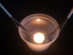
I placed a lighted candle into a capillary bed at the bottom of the illustration to represent fuel burning aerobically in a single cell.
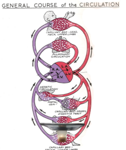
The image below illustrates both the big picture of general circulation and the microscopic areas.
The largest blood vessel of the human body is the aorta. It stems directly off the heart and then branches into progressively smaller arteries, and arteries get smaller and smaller until they become capillaries, which are invisible to the naked eye. The finer tendrils of roots are like capillaries; each are the ‘thinnest tubes’ through which water, blood, and nutrients flow.
The microscopic space of your cardiovascular system is called a capillary bed – where ‘who you are microscopically’ exists as a collection of cells collected tightly together in organized specific structures called tissues. Specific cells make specific tissues or body parts, e.g. brain, kidney, lungs, muscles, etc.
This next image shows individual cells – clearly labeled as cells/tissues – embedded within a capillary bed.
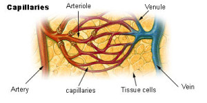 This is the area where the ‘exchange’ of gases, nutrients, and waste products happens. In class, I had you draw small tiny circles within one of the capillary beds to drive this point home: circles represent cells.
NOTE: Learning super advanced concepts in the future are made simple by using circles to represent cells.
I showed a more elaborate model called The Straw Prop – along with a lighted candle to explain more stuff. Click to enlarge the image.
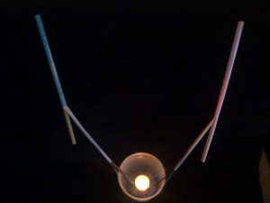 The Straw Prop represents the entire circulation system from where blood, oxygen, and nutrients exit the heart. Then after a cell burns fuel, blood flows back to the heart with CO2 rich blood and other waste products.
The largest red straw represents the aorta coming directly out of the left ventricle of the heart. The tiny thin straws represent the capillaries, and the candle represents mitochondria of a cell. Red straws represent arteries, which are rich in O2. Blue straws represent veins or the venous system, which have less O2 and more CO2 as a result of burning fuel in cells.
The mitochondria is ‘the space’ or section of a cell where O2 combines with fuel, just like a firebox of a coal burning locomotive or Weber grill. The candle represents mitochondria.
The formula for respiration or aerobic or oxidative metabolism, which I expect you all to know by heart is: Fuel + O2 –> CO2 + H2O + Heat.
Again, the microscopic cellular space is the energetic ‘alive’ region of the body that makes us ‘who we are’. Following up from last week – this is the area of the body we actually want to ‘cleanse’ – since we want to maintain a clean, watery, properly balanced electrolytic environment surrounding our cells. Right? Whoever thought it was so simple? Just kidding.
Anyway, this explains why I used a mosquito ‘bite’ to describe ‘interruption of transport of materials’ – by way of destroying or ‘blowing up capillaries’ – which results in swelling you see on your skin. I didn’t mention this in class, but histamine is ‘the bomb’ which destroys the capillaries so that blood transport of the anticoagulant protein injected by the mosquito can’t circulate. This swelling is only one form of inflammation, which I will describe in more specific ways later.
FYI: I may use the straw prop or the black and white illustration of ‘general circulation’ to test you and identify/label the following parts.
- Heart
- Artery or Arterial System
- Vein or Venous System
- Mitochondria
- Label what section contains more or less oxygen or CO2.
- Capillary bed
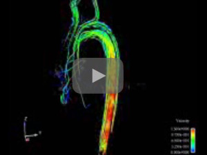
Here’s the 4D MRI of blood flowing out of the heart. This file was corrupt and I couldn’t show it yesterday.
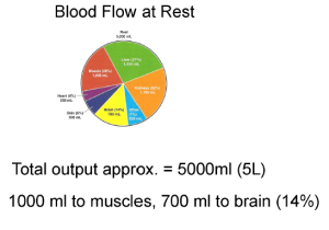
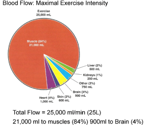
Since blood fills this system up, I showed the two pie charts to visualize the difference in distribution of blood flow at rest and during maximal intensity exercise.
On average – people have about 5 liters total blood in their blood vessels – including the heart, arteries, capillaries, and veins. The left figure shows the volume of blood pumped in 1 min at rest – approximately 5 liters. The right figure shows the volume of blood pumped in 1 minute during max exercise intensity. It went from 5 liters/min to 25 l/min.
In a 100ml wad of blood – freshly oxygenated just after leaving the heart – there is always about 20 ml of O2 dissolved in blood as gas. Recall, oxygen dissolves as gas in blood or water. Cold water allows more O2 to dissolve in it, which is why there is more life on cold ocean water than tropical sea water. Oxygenated gasoline has O2 dissolved in it, which is why it screws up small engine carburetors; we’ll examine why this important in terms of health, performance, and nutrition later.
At rest – less O2 is used compared to any greater intensity level of exercise. So within a 100ml wad of blood coming back to the heart, we’ll see about 12ml O2 left over. During maximal exercise, we’ll see about 4ml O2 remaining. (Meaning we extracted 16ml during max exercise out of a wad of blood)
I will ask the following question verbatim on the test:
- Why is there less O2 in the venous system compared to the arteries?
Understanding the O2 graph.
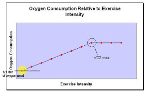 The horizontal x-axis shows exercise intensity level from rest to max efforts. The extremes of human movement are ‘at rest’ or ‘sprinting’. So we could label the x axis on the left ‘rest’ and on the right ‘high’ intensity. Or you could label the horizontal ‘x’ axis more specifically in terms of familiar movements or even images, e.g. sitting, walking, biking slowly, rock climbing, biking quickly, or sprinting.
The yellow shaded dot marks the volume of O2 combining with fuel while resting – which is around around 1/3 liter used on the average by a human. Each of us consumes a slightly different amount at rest – but whatever this amount for anybody at rest is – is called 1 Met – which stands for metabolic equivalent. If you are DEAD you consume no oxygen. So ‘zero Mets = the metabolic equivalent of being dead where you consume zero oxygen, whereas 1 met is equal to the amount of oxygen consumed at rest or ‘idle’.
3 mets would be 3 times the amount of O2 used at rest, so this would be 3 times 1/3 a liter of O2, which equals 1 liter.
The vertical ‘y’ axis represents O2 consumed, which obviously increases as you work harder or speed up. For example, at 2 liters per minute you’d definitely be moving and working.
Some folks started drawing the line in the bottom left hand corner – which indicates zero oxygen consumption – and so in class I said this means, “you are dead”. Do not write the graph that way.
Most people think in term of ‘maximizing oxygen uptake’ – but I turned this around and told everyone to think about what happens if your level of oxygen consumption at rest drops from its ‘normal’ 1 Met level. I called this the bottom dropping out.
The image below refers to people who need oxygen services if their ‘bottom drops out’.
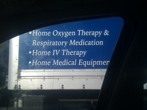 People needing this service have trouble getting O2 into their cells for some reason at rest. I took this picture through my passenger side window while stopped at a red light in 2012.
Perhaps a better marker of true health as you age is the ability to keep using whatever your 1 met is. This means you must maintain the ENTIRE system – e.g. your heart, blood vessels, blood itself, lungs, – so your cells can maintain using the same amount of oxygen at rest forever. This means you have not diminished at the low end of oxygen consumption – or that your body at rest is healthy – and your 1 met 40 years from now is nearly the same as it is today. (I will ask why this is important on the test)
From this point of ‘healthy cells at rest’ exercise only speeds up from what in the beginning is already operating the way it should. I mentioned this provides a better way to understand strokes and cognitive decline. A couple people immediately connected these concepts to effects experienced from sleep apnea and cluster headaches.
If you are compromised at rest then it’s probable that speeding up – you may still be compromised, it’s difficult to work harder and it could even cause harm. People who need oxygen at rest obviously have trouble somewhere within the system – with one part or some parts, if not all the parts – all the way down to the cell level. So they may not use the proverbial 1/3 liter of oxygen or ‘1 met’ for some reason.
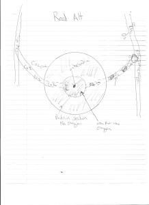
Using blank bar coasters we drew the ‘exchange of energy and matter’ at the cell level similar to this image below:
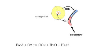 Exchange and transformation of energy and matter indicates a cell is living or the chemical formula for aerobic metabolism is happening.
This is a strange concept at first…. your blood stream never touches ‘who you are’ – meaning capillaries never contact your cells. Notice there is space between the cell and the capillary. Oxygen gas diffuses out of a capillary along with nutrients and move into a cell. Then CO2 moves out and back into the capillary. I said I want you to visualize the space and draw it. All this requires is drawing a circle and a single capillary, so don’t get lazy and fail to get good at this!
I also explained at the start of lecture – energy is NOT created – it only transforms. We are a conduit for energy; we transform the energy ultimately from the sun, into stored matter (mass), motion, or heat. Are we not spiritual beings having a human experience? That’s something to ponder about on our own or talk about over drinks with friends, no?
Anyway…
Here’s another rendition of the exchange of energy and matter drawn by a 2012 student using the bar coaster – click to enlarge:
Images from Body Worlds. The weird looking pic shows the vessels from the small intestine – specifically the jejunum.
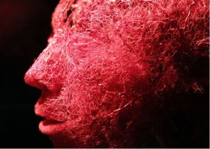
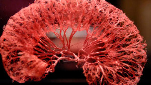 As I said yesterday, YOU DO NOT SEE capillaries, what you see are arteries and arterioles. Capillaries and cells are not seen by the naked eye.
I showed all this to get you to develop ‘spatial sense’ – specifically to see the SPACES of the body in a more complete way and realize the action of life, nutrition, and exercise is happening at a level where we do not see it. We see and feel the expression of it when we move and touch others and feel their body heat. This space we do not see – the microscopic space – is the space we want to ‘cleanse’ in terms of cleansing the body. This means you must have quality blood, and excellent blood flow within capillaries in order to deliver nutrients AND remove waste products, and lastly healthy kidneys!
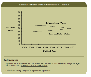
Finally, I showed this image:
This shows the idea dehydration is: less water resides within cells. (Intracellular water decreases). The body gets ‘leaky’ as we age or have crappy nutrition. I gave a very graphic description of plasma leaking out of the skin yesterday; a severe case of lymph edema. Capillaries in the body are designed to ‘leak’ somewhat or not due to how tightly the cells that make the wall are packed together.
Older people have less water in their bodies, which means less water in cells. Drinking water may not hydrate you as well as you think; water balance is maintained electrically, i.e. by electrolytes. More on this later.
Update:
At the beginning of this lecture I was asked when we do some physical stuff. I answered and then explain before the class “We are moving from understanding the big picture – developing visual/spatial sense of the whole – then today I am detailing the connection to the small picture, i.e. what happens in the cells. This is so you know what you are actually doing with your body and can decisively and persuasively tell people how the body works.
Immediately, I demonstrated what I meant based on the idea, “There is no such thing as stretching a muscle” – showing my bicep working – muscles lengthen or contract – what you feel as ‘the stretch’ is – described and demoed with Whitney doing the hand clasp, Ashley doing push hands/leverage, and Greg as the volunteer to do the splits and .
- You hear ‘actin and myosin’ slide – cell level stuff – but let’s make sense of it at the cell level and the whole level at the same time! (Hand clasp demo)
- Explained leverage and counter force with Ashley and used the door to get the point home.
- Greg demoed the 1-leg up – no stretch. Then I went on from here to explain the physics.
LINKS:

